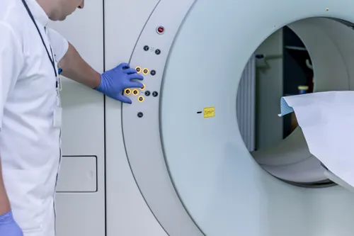
The type of imaging ordered by your doctor will depend upon your injuries.
- X-Ray: An X-ray, also called a radiograph, sends radiation through the body which is blocked by tissues containing calcium (bones and teeth), but passes through the body’s soft tissues, producing a contrasted image.
- CT Scan: A computerized tomography (CT) scan takes a series of X-ray images from all angles to produce detailed images of the body’s soft tissues, like tendons, ligaments and venous structures.
- MRI: Magnetic resonance imaging (MRI) uses tube-shaped magnets which work by temporarily realigning water molecules in the body. Radio waves cause these aligned atoms to produce faint signals, which are used to create cross-sectional MRI images — like slices in a loaf of bread.
- DTI: Diffusion tensor imaging tractography, or DTI tractography, is an MRI technique that measures the rate of water diffusion between cells to map the body’s internal structures and is commonly used to provide images of the brain.

